


WHAT ARE MIGRASOMES
What are migrasomes?
Specialised organelles involved in intercellular communication
The review describes migrasomes as specialised organelles involved in intercellular communication. They were first identified in migrating cells: as a cell moves, it leaves retraction fibres, and migrasomes commonly form at fibre junctions or tips.
While tethered to the cell, a migrasome can act as a local platform for exocytosis; after detachment, it has the properties of a large extracellular vesicle. Typical diameters are about 500–3000 nm, and the lumen can contain multiple smaller vesicles.
Migrasomes are not confined to culture dishes. The review brings together ex vivo and in vivo observations from zebrafish embryos, the chicken chorioallantoic membrane, and mouse vasculature, liver, and spleen.
Distinguishing Migrasomes from Other EVs
Migrasomes vs conventional extracellular vesicles
| Five vesicle types | 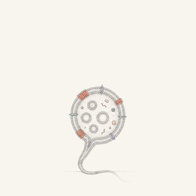 |  | 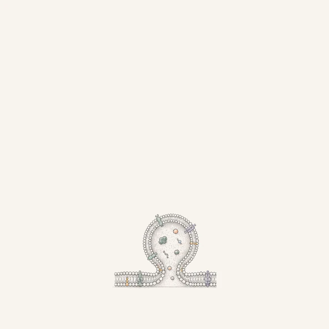 | 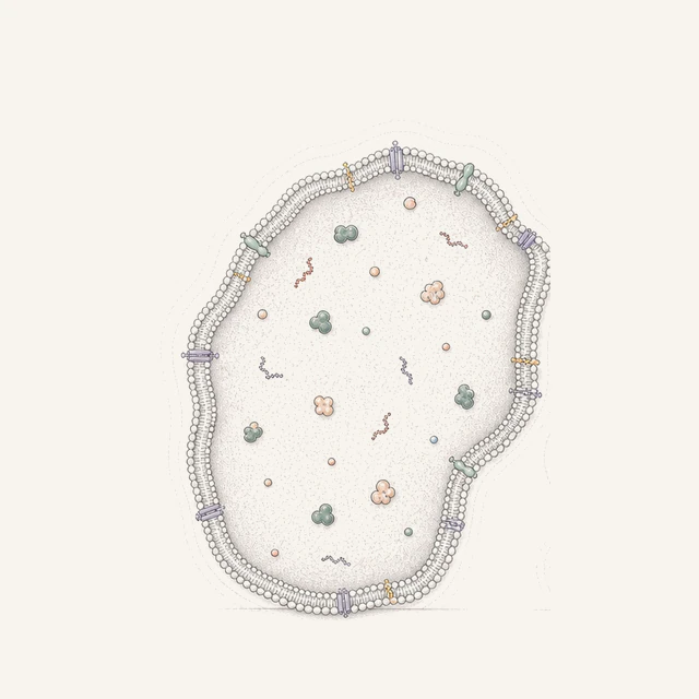 | 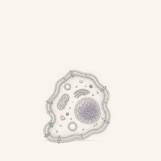 |
|---|---|---|---|---|---|
| Property | Migrasomes | Exosomes | Microvesicles | Large oncosomes | Apoptotic body |
| Location | Tethered to migrating cells; large extracellular vesicles after detachment | Extracellular and separate from the cell body | Extracellular and separate from the cell body | Released extracellularly by tumour cells | Released extracellularly by apoptotic cells |
| Size | 0.5–3 μm | 35–150 nm | 0.1–1 μm | 1–10 μm | 1–5 μm |
| Formation | Retraction-fibre derived and Tspan4-dependent | Multivesicular body (MVB) pathway | Plasma-membrane budding | Tumour-cell membrane blebbing and shedding | Apoptosis |
| Canonical markers | ITGs, Tspan4 | Alix, CD63, Tsg101 | ARF6, KIF23 | ARF6, Caveolin-1, GAPDH | cCaspase-3, PS |
| Biological roles | Directed cell migration, short- and long-range communication, organ development, mitochondrial quality control, and coagulation | Long-range cell communication, immune regulation, tumour-microenvironment control, and targeted delivery | Coagulation, inflammatory signalling, angiogenesis, and tumour-microenvironment remodelling | Transfer of tumour-associated cargo that promotes invasion, metastasis, and microenvironment remodelling | Induction of macrophage engulfment while limiting severe inflammation |
| Research value | Tumour metastasis and microenvironment; cardiovascular, cerebrovascular, and inflammatory disease; liquid-biopsy biomarkers; complex-drug delivery | Liquid-biopsy biomarkers and natural drug-delivery systems | Cardiovascular and coagulation mechanisms; disease-activity and acute-injury monitoring | Tumour progression and metastasis; microenvironment and resistance mechanisms; tumour biomarkers | Apoptosis pathways and mechanisms; immune tolerance and autoimmune disease |
Migrasome cargo
Migrasomes carry diverse information and materials inside and out
The review classifies migrasome cargo into five categories: secretory vesicles, damaged mitochondria, mRNA, viruses, and coagulation factors.
Secretory vesicles, damaged mitochondria, virus particles, and mRNA reside in the lumen, whereas coagulation factors on neutrophil-derived migrasomes are adsorbed to the outer surface. These locations are linked to different loading and release routes, some of which remain unresolved.
Yu & Yu, Cell Research (2025), Fig. 3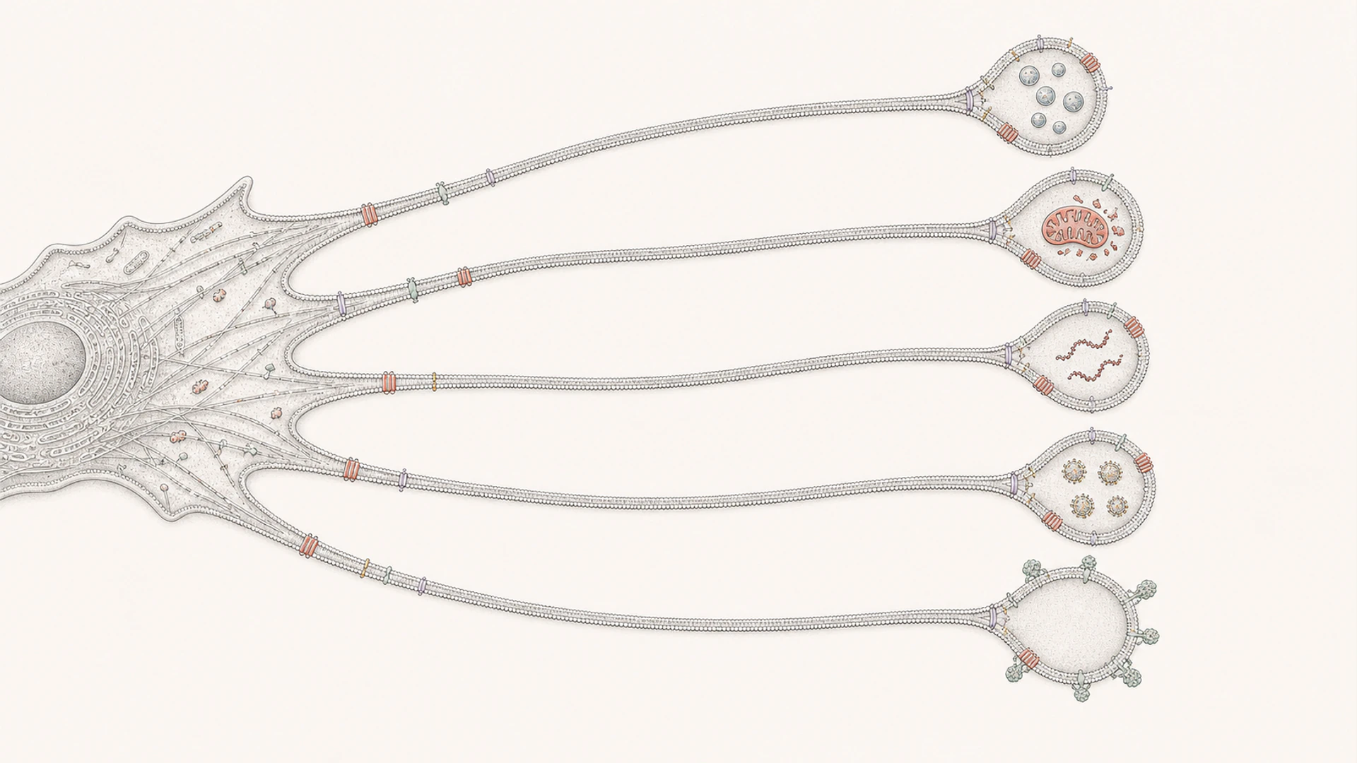
- Secretory vesicles
- Damaged mitochondria
- mRNA
- Viruses
- Coagulation factors