


1 / 4
MigraSee®︎ Migrasome and Submicron-to-Micron Particle Observation Chip
SE001
For high-magnification observation and counting of 0.3-10 μm vesicles or particles, using a fixed 20 μm channel height to control the confocal z-stack volume.
 Product manual
Use this page for product selection and experiment planning. Consult the SE001 series manual for the full procedure and limitations.
MigraSee®︎ supports high-magnification observation and counting of vesicles or particles spanning the micron scale. Biological fluorescence labeling enables the morphology of specific targets in mixed samples to be observed and their concentration to be estimated.
The chip uses a fixed channel geometry to define the confocal z-stack volume and calculates concentration from the particle count within that volume, supporting efficient, convenient observation.
Components and storage
| Component | Qty. | Storage |
|---|---|---|
| MigraSee chip | 42 single-use loading channels | Store as directed in the product manual. |
| Plasma sample stabilizer | 100 μL | Store as directed in the product manual. |
| Product manual | 1 copy | Store as directed in the product manual. |
| MigraSee®︎ product file package | 1 package | Store as directed in the product manual. |
PRODUCT FEATURES
Product advantages
Designed for migrasomes
Optimized for the approximately 0.5–3 μm vesicular structure of migrasomes to support efficient, convenient observation.
Concentration measurements across the micron scale
Supports observation and concentration measurement of migrasomes and similar particles spanning the micron scale, extending beyond the limitations of conventional particle measurement.
High-throughput quantitative counting
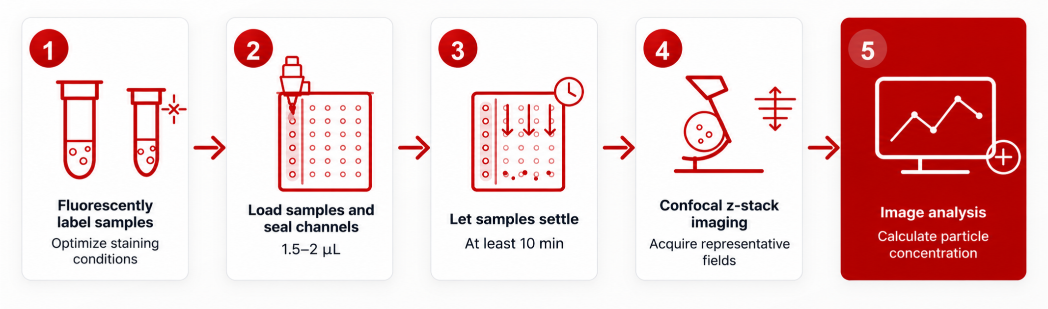
Requires only 1.5–2 μL per sample and supports 42 tests per chip for batch observation and analysis.
Precise concentration and morphology characterization
A fixed channel height precisely defines the confocal z-stack imaging volume for particle counting and absolute concentration calculations.
EXPERIMENTAL WORKFLOW
Workflow
EXAMPLE RESULTS
Example results
1 / 1
Counts of neutrophil-derived Ly6G+ migrasomes in blood from bacteria-injected and uninjected mice using this chip (left) are broadly consistent with the imaging-flow-cytometry counts reported by (Jiang et al., Nat Cell Biol, 2024) (right).
FAQ
Frequently asked questions
What is the SE001 channel height?
The confirmed channel height is 20 μm. The z-stack should cover the full channel plus any necessary margins to avoid missing particles near its upper or lower boundaries.
How many samples can one chip process?
Each chip contains 42 single-use loading channels. Mark a channel immediately after use to prevent reloading.
Why might particle counts be unstable?
Fluorescence background, particle drift, unsuitable concentration, incomplete z-stack coverage, and threshold selection all affect counting. A loading protein concentration of 100 to 200 µg/mL by BCA is recommended. For a new sample, first assess settling of the undiluted sample, then test serial dilutions if it is too concentrated and optimize settling time and imaging settings.
How can the channel base be located quickly?
In bright field, align the objective with the channel and adjust focus along z until the channel boundaries are sharp. Use the central markers to adjust x and y, then switch to the fluorescence channel to locate the sample.
How can z-stack acquisition be made more efficient?
Choose the largest z-step and an appropriate stack height that still support the target-size analysis. A microscope with an automated stage can batch-acquire multiple channels and positions.
Manual and COA
RELATED PRODUCTS


