


How to study migrasomes
Migrasome research methods
From observing migrasomes to understanding their functions and mechanisms
Migrasome research can follow five stages: observation, isolation, identification, manipulation, and studies of function and mechanism. Each addresses a different experimental question, from establishing where migrasomes form and how they are distributed, to obtaining samples, changing their number or composition, and testing their effects on cells and tissues.
Download research handbook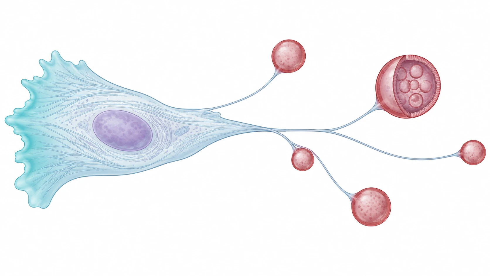
- Observation
- Isolation
- Identification
- Manipulation
- Function and mechanism

01Observation
See where migrasomes form and how they are distributed
Migrasomes form on retraction fibres left behind by migrating cells. Three labelling approaches can be used: fluorescent marker fusion proteins, dyes and fluorescent probes, and fluorophore-conjugated antibodies. Microscopy then records their location, formation and changes in number.
- Fluorescent marker fusion proteins
- TSPAN4-GFP or TSPAN4-mCherry, for example, can reveal localisation and formation in live cells.
- Dyes and fluorescent probes
- Choose fluorescent WGA, FM4-64 or Calcein-AM according to the sample and imaging requirements.
- Fluorophore-conjugated antibodies
- Use validated antigens to identify migrasomes or their cell of origin, distinguishing fixed-sample staining from surface labelling in vivo.
Fluorescent marker fusion proteins: observing formation and localisation
Huang and Yu's 2024 methods article describes three fluorescent labelling strategies. The first expresses fusion proteins such as TSPAN4-GFP and TSPAN4-mCherry in cells to visualise migrasomes and their connections to retraction fibres, including by live-cell time-lapse imaging.
The figure shows TSPAN4-GFP-labelled L929 cells at different focal planes. Fusion protein expression levels may affect migrasome formation, so experiments should account for expression levels and include appropriate controls.

Dyes and fluorescent probes: rapid migrasome labelling
Huang and Yu show WGA555-stained NRK cells, comparing red WGA signals with green TSPAN4-GFP. Dyes such as FM4-64 and Calcein-AM can also be considered according to the study. Live-cell suitability, fixed-sample use and photobleaching characteristics need separate consideration.
WGA is a fluorophore-conjugated lectin, not a migrasome-specific antibody. Interpret staining alongside connections to retraction fibres and migrasome morphology.
Huang & Yu, Biophysics Reports, 2024 · Fig. 3, Table 3 ↗
Fluorophore-conjugated antibodies: identifying endogenous surface markers
Jiang et al. labelled mouse neutrophils and their migrasomes with PE-conjugated anti-Ly6G antibodies, while AF647-WGA labelled blood vessels, to observe migrasome formation by intravital imaging. This example shows how a validated surface antibody can be used for in vivo observation.
Fixed samples can also use a primary antibody with a fluorescent secondary antibody; Huang and Yu's Fig. 1 and 2 show ITGA5 and ITGB1 immunofluorescence. Distinguish directly conjugated primary antibodies from indirect secondary-antibody labelling, and check the species, antigen and sample conditions.
Jiang et al., Nature Cell Biology, 2024 · Fig. 1b ↗Huang & Yu, Biophysics Reports, 2024 · Fig. 1, 2 ↗
Collect together → Differential centrifugation → Purification
Crude fraction → Immunomagnetic enrichment
Pretreatment → WGA bead enrichment
Mince/digest → Remove cell bodies → Purify
Choose the route for the sample, then verify identity and sample quality after isolation
02Isolation
Obtain samples for subsequent studies
Isolate and enrich migrasomes from cultured cells, blood, urine or tissues for compositional analysis and functional experiments. The method must balance recovery, purity and structural integrity while accounting for the characteristics of each sample type.
- Differential centrifugation
- Remove cells and larger debris to obtain a crude migrasome fraction.
- Density-gradient centrifugation
- Separate fractions further and reduce co-isolated contaminants.
- Affinity capture
- Enrich target fractions through surface-molecule binding in sample systems where the approach has been validated.
Blood study: isolating migrasomes with immunomagnetic beads
Jiang et al. (Nature Cell Biology, 2024) isolated neutrophil-derived migrasomes from mouse blood. In platelet-depleted mice, they removed blood cells by centrifugation, collected a crude fraction, and then used anti-Ly6G-conjugated magnetic beads for positive capture. The paper also compared purification using a neutrophil negative-selection kit.
Scanning electron microscopy showed bead-coated migrasomes with connected retraction fibres. Ly6G capture applies to this mouse-derived system. A different species or cell of origin requires selection and validation of an appropriate capture marker.

Optional tools: use Migrasome Therapeutics migrasome isolation kits, selecting a protocol for the specific sample type.
Kits provide an optional product-based route; the Jiang paper used the immunomagnetic methods described above.
Cultured cells: recovering migrasomes that remain attached
The Li Yu Lab protocol collects cultured cells together with attached migrasomes, removes cells and larger debris by differential centrifugation, then uses density-gradient purification. Collecting only the culture supernatant may miss migrasomes that remain attached to retraction fibres or the culture dish.
Optional tools: use Migrasome Therapeutics migrasome isolation kits, selecting a protocol for the specific sample type.
- Collect together
- Differential centrifugation
Urine: WGA bead enrichment
Yang et al. combined WGA bead enrichment with flow cytometry and analysed markers of podocyte origin. The identity and origin of the target particles still require verification after capture.
- Sample pretreatment
- WGA bead enrichment
- Flow cytometry and origin verification
Tissues: release by digestion, then purify
Mince and enzymatically digest the tissue, remove cell bodies by differential centrifugation, then purify by a density gradient or a Migrasome Therapeutics kit. Adapt digestion conditions to the tissue type and check migrasome structure and markers after isolation.
Optional tools: use Migrasome Therapeutics migrasome isolation kits, selecting a protocol for the specific sample type.
- Mince tissue
- Enzymatic digestion
- Remove cell bodies by differential centrifugation
Fibre connections
Membrane and internal vesicles
NDST1 · PIGK
CPQ · EOGT
Source cells
Other vesicles and contaminants
Morphological and molecular evidence together support identification
03Identification
Confirm migrasomes in the sample
Identification combines evidence of origin, morphology and molecular composition. Microscopy reveals structural features, panels of markers support identification, and analysis of co-isolated components helps assess sample quality.
- Fluorescence and electron microscopy
- Examine connections to retraction fibres, membrane structures and internal morphology.
- Immunofluorescence and Western blotting
- Detect migrasome-associated proteins and assess their enrichment.
- Analysis of control fractions
- Assess co-isolated components, including other vesicles, cell debris and soluble proteins.
Study example: interpreting electron microscopy alongside protein analysis
Zhao et al. combined electron microscopy of purified fractions with protein analysis and proposed migrasome-enriched markers including NDST1, PIGK, CPQ and EOGT. Similar designs can compare candidate samples with their source cells and other vesicle fractions.
These markers require validation in the specific sample system. A single positive band or a particle-size range alone is insufficient to identify migrasomes.


Freshly isolated samples: checking PS exposure with Annexin V
Annexin V recognises phosphatidylserine (PS) exposed on the outer membrane surface. Jiang et al. tested purified blood neutrophil-derived migrasomes and found that most did not expose PS, whereas the neutrophil microparticles (NMPs) used for comparison showed a clear Annexin V signal.
The figure retains the original distributions for the unstained negative control, NMP control and migrasomes. For freshly isolated migrasomes, absent PS exposure or low Annexin V signal can serve as a supporting feature alongside morphology and a marker panel. Annexin V negativity alone cannot establish identity, and the result should not be assumed to apply to every source or processing condition.

Normal expression
Inhibit a formation-promoting gene
Restore target gene expression
Monitor cell viability, migration and retraction fibres in parallel.
04Manipulation
Change migrasome number or composition
Genetic interventions, changes in culture conditions and compound treatments can help investigate migrasome formation and establish comparisons for functional experiments. Cell viability, migration and retraction fibres should be monitored at the same time.
- Gene overexpression, knockdown or knockout
- Test how candidate molecules affect migrasome formation or composition.
- Substrate changes and compound treatment
- Identify conditions or pathways that affect migrasome formation.
- Genetic rescue
- Test whether restoring the target molecule reverses migrasome-related changes.
Study example: investigating ROCK1 through compound screening and genetic intervention
Lu et al. used fluorescence imaging to screen compounds that affect migrasome formation, followed by ROCK1 knockdown for further validation. They counted migrasomes both per cell and per unit length of retraction fibre, helping distinguish reduced migrasome formation from a reduction in retraction fibres.
The same approach can be used to study candidates such as TSPAN4. Drug effects require validation through genetic experiments and should not be equated directly with migrasome-specific inhibition.

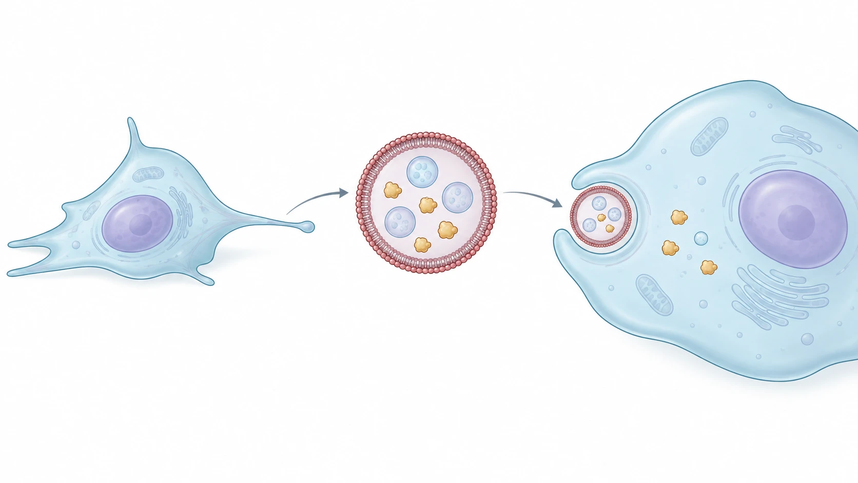
Combine cargo interventions with functional assays
Test key molecules and pathways and their effects on cell behaviour.
05Function and mechanism
Determine what migrasomes do and how they act
After confirming identity and establishing methods of manipulation, test how migrasomes affect cell behaviour, tissue function or disease processes, then investigate the key molecules and signalling pathways involved. Functional experiments measure effects; mechanistic experiments explain how those effects arise.
- Formation interventions and migrasome supplementation
- Test whether migrasomes contribute to a biological effect.
- Uptake tracing and functional assays
- Measure changes in proliferation, migration, angiogenesis and other readouts after recipient cells contact or take up migrasomes.
- Omics screening and molecular validation
- Identify candidate cargo, then test its role through cargo knockout, receptor blockade or pathway intervention.
Zebrafish development: comparing migrasome deficiency and supplementation
Jiang et al. (Nature Cell Biology, 2019) found that migrasomes in zebrafish gastrulae carry chemotactic signals and contribute to the correct positioning of dorsal forerunner cells (DFCs) and organ morphogenesis. Migrasome formation is impaired in MZtspan4a and MZtspan7 mutants.
The study injected PBS or migrasomes into wild-type embryos and both mutant types, then measured Kupffer's vesicle (KV) size and cilia number at the six-somite stage. Comparing deficient and supplemented groups tests whether migrasome supplementation can improve the developmental phenotype. Analysis of chemotactic signals helps explain the mechanism.
01Establish control and migrasome-supplemented groups
Normal development reference
Impaired migrasome formation
Test for phenotype improvement
02Examine Kupffer's vesicle (KV) at higher magnification
- Vesicle size
- Compare normal KV formation
- Cilia number
- Compare cilia development within KV
Deficiency effectMutant + PBSversusWild type + PBS
Supplementation effectMutant + migrasomesversusMutant + PBS
Coagulation: can supplementation restore haemostasis after migrasome reduction?
Jiang et al. reduced neutrophil-derived migrasomes through neutrophil depletion and genetic intervention in Tspan9, then assessed coagulation using tail-tip bleeding, platelet aggregation and imaging at injury sites.
Fig. 5q and r compare mice with conditional Tspan9 deletion, controls, and mice supplemented with purified migrasomes after deletion. Supplementation reduced blood loss, supporting a role for migrasomes in haemostasis. This design connects a deficiency-associated phenotype with recovery after supplementation. Neutrophil depletion also affects the cells themselves, so genetic models and multiple functional readouts are needed to support the interpretation.
Jiang et al., Nature Cell Biology, 2024 · Fig. 5i, j, q, r ↗
PTEN: identifying the cargo molecule responsible
Zhu et al. added L929-cell-derived migrasomes to PTEN-deficient recipient cells, comparing migrasomes from normal and Pten-knockout donors. Migrasomes from normal donors inhibited AKT signalling and proliferation more strongly, supporting a contribution from PTEN cargo.
Alongside deficiency and supplementation experiments, cargo knockout can identify which migrasome component contributes to the effect.
Zhu et al., Cell Research, 2021 · Fig. 1j, n, o ↗
Migrasome extraction and purification tools by sample
These are optional product workflows from Migrasome Therapeutics. They are distinct from the methods used in the papers above; follow the relevant product manual for product-specific procedures.
- Blood samples
Select extraction kits by species and total or specific cell source.
- Cultured cells
Compare complete production, crude isolation and purification workflows using density gradients or magnetic negative selection.
- Tissue samples
Begin with a user-prepared tissue single-cell suspension, followed by crude isolation and magnetic negative selection.
RESEARCH TOOLS HANDBOOK
Migrasome Research Tools Handbook
Explore migrasome research methods and tools for isolation, purification, imaging, and counting.