


Formation Mechanisms
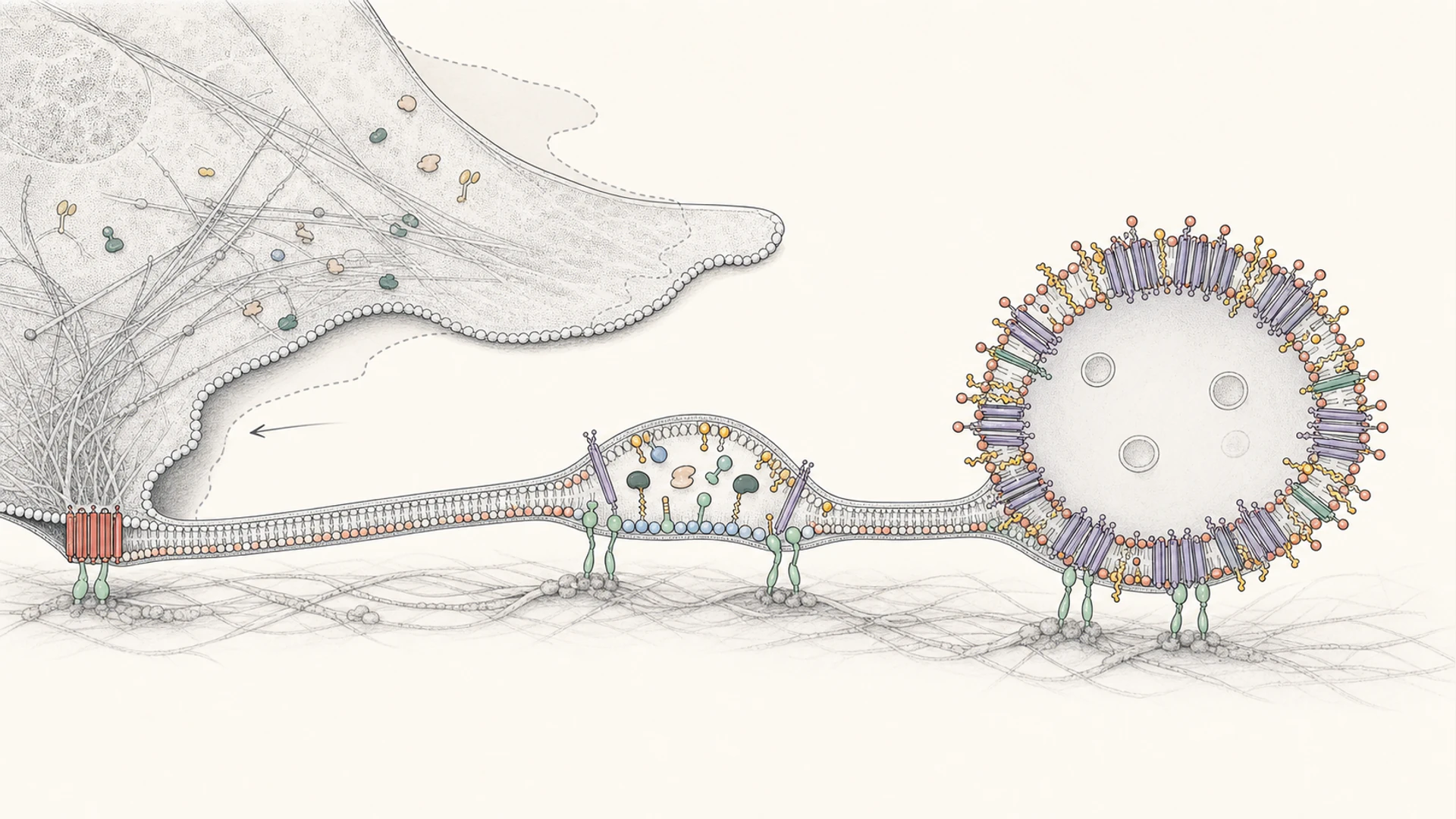
Mechanistic analysis of migrasome formation
Formation site
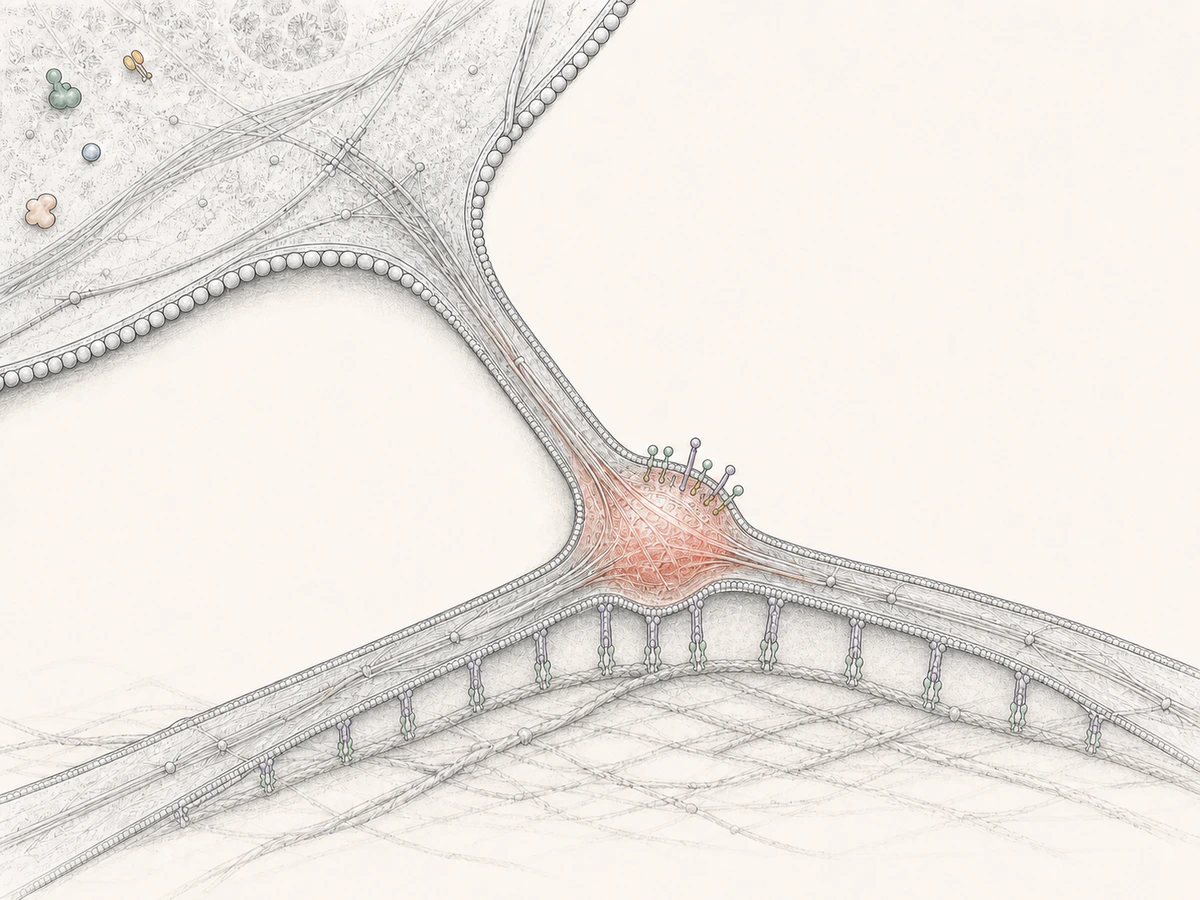
Migrasomes enlarge on retraction fibres left behind by a migrating cell rather than at arbitrary positions on the cell body. Substrate contact and fibre geometry constrain early growth.
Relative-displacement dependence
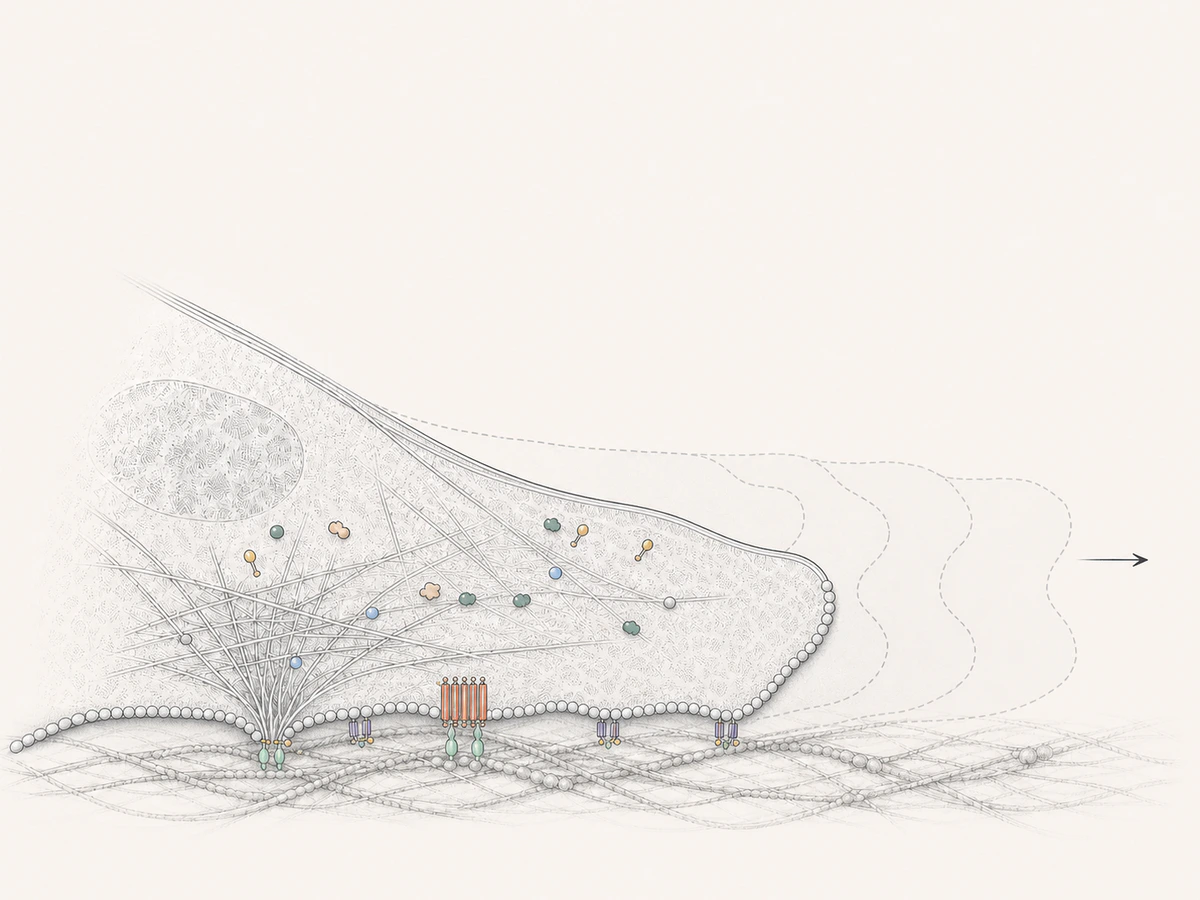
As the cell moves forward, SMS2 foci anchored at its basal leading edge remain on the substrate and come to lie on the trailing retraction fibres. Formation therefore depends in part on movement of the cell relative to the substrate.
Membrane origin and cargo
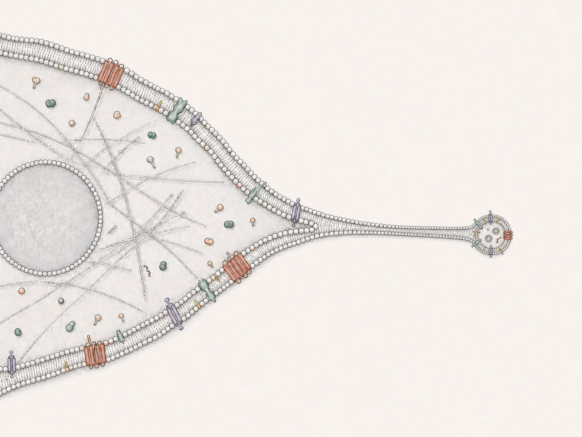
Retraction fibres form a material-transport route between migrasomes and cells, providing a continuous source of plasma membrane and luminal contents. Proteins, nucleic acids, organelles, and secretory vesicles enter migrasomes through distinct transport and sorting mechanisms.
Stages of Formation
Migrasome Formation: From Nucleation to Maturation
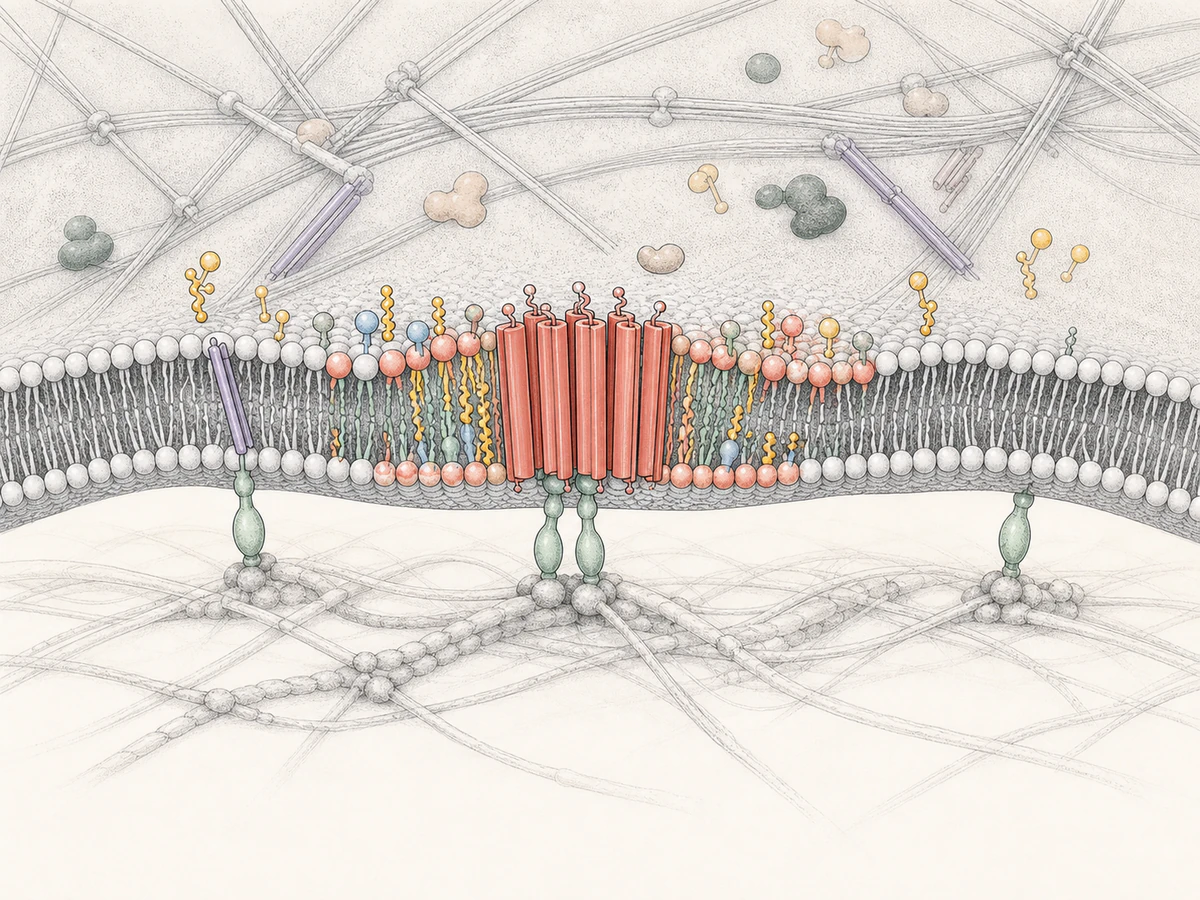
SMS2 nucleation
At the basal leading edge, discrete SMS2 foci produce sphingomyelin and stay anchored to the substrate. Cell movement leaves these lipid sites on the retraction fibres.
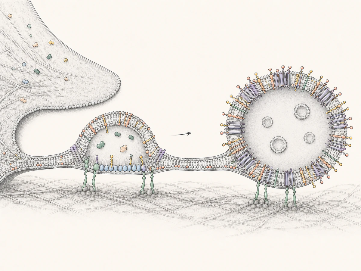
Lipid- and integrin-mediated migrasome maturation
PIP5K1A produces PI(4,5)P2, which recruits Rab35. Rab35 then binds the GFFKR motif of integrin α5, establishing an extracellular-matrix-facing anchoring axis.
At this stage, the migrasome formally forms and begins to grow.
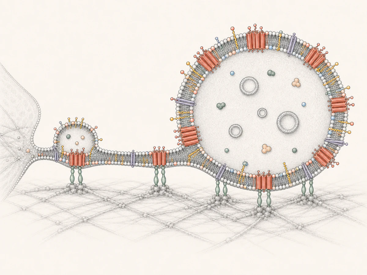
TEM-mediated membrane expansion and stabilization
Tension fluctuations and retraction-fibre junctions promote membrane bulging, while Syt1 can stabilize early precursors in a calcium-dependent manner. TSPAN4 and other tetraspanins then co-assemble with cholesterol and sphingomyelin in the membrane to form TSPAN4-enriched microdomains (TEMs). TEMs further self-organize into TSPAN4-enriched macrodomains (TEMAs), continuously increasing local membrane bending rigidity, promoting structural expansion, and ultimately generating migrasome-like structures.