


Cancer Biology
- Tumor microenvironment
- Metastasis-associated communication
- Immune modulation
MIGRASOME SCIENCE
Migrasomes are specialized organelles generated during cell migration that enable spatially controlled signaling, molecular transfer and cellular homeostasis.

01 / WHAT
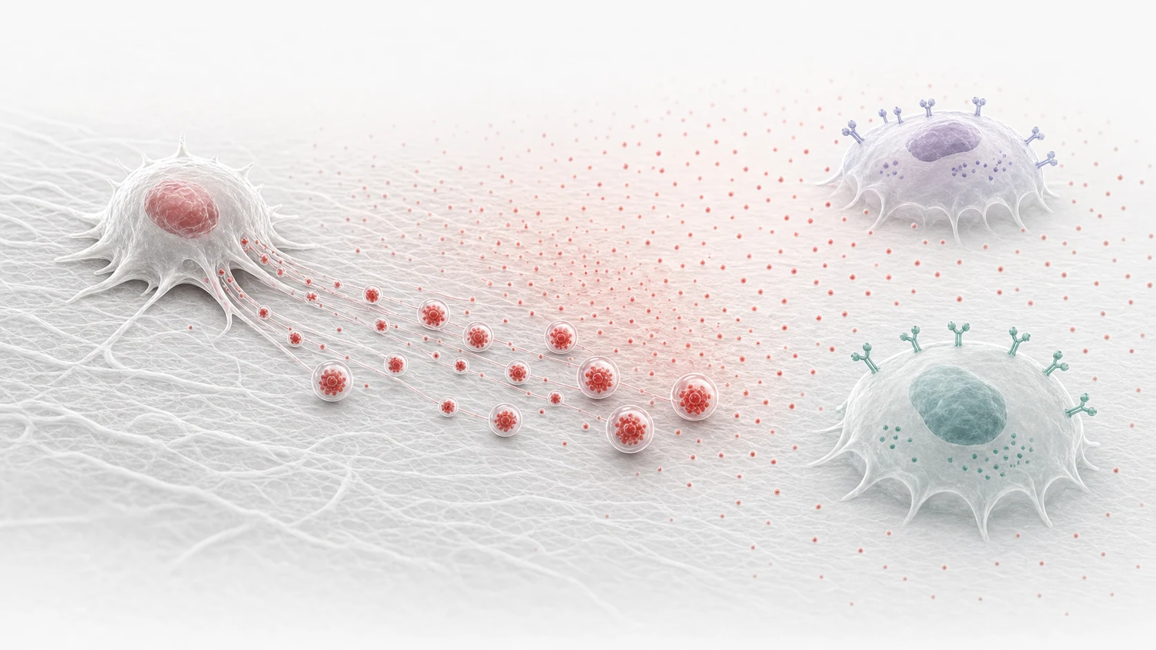
Unlike conventional extracellular vesicles, migrasomes arise through a migration-dependent process and show distinctive features including retraction-fiber connection, a large membrane-bound architecture, intraluminal vesicles and enriched molecular cargo.
Cell migration is fundamental to development, immunity, tissue repair and disease progression. Migrating cells leave behind membrane structures called retraction fibers, where migrasomes form and reveal a previously unknown connection between cell movement and intercellular communication.
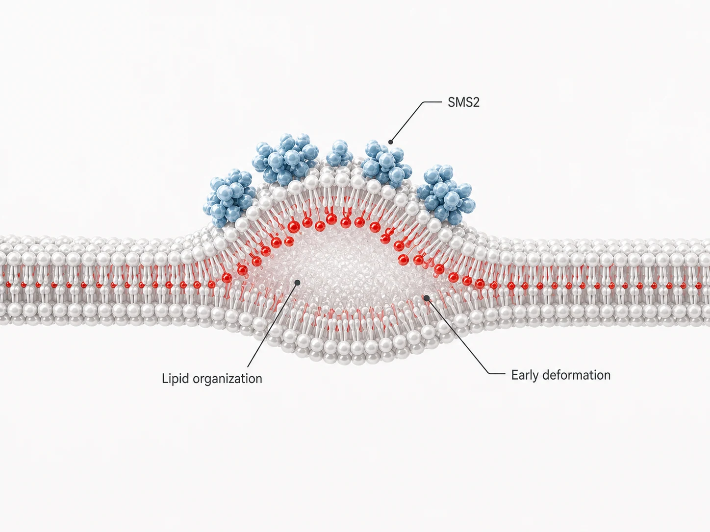
Understand what migrasomes areSMS2-dependent sphingomyelin production defines specialized membrane domains that initiate migrasome formation.
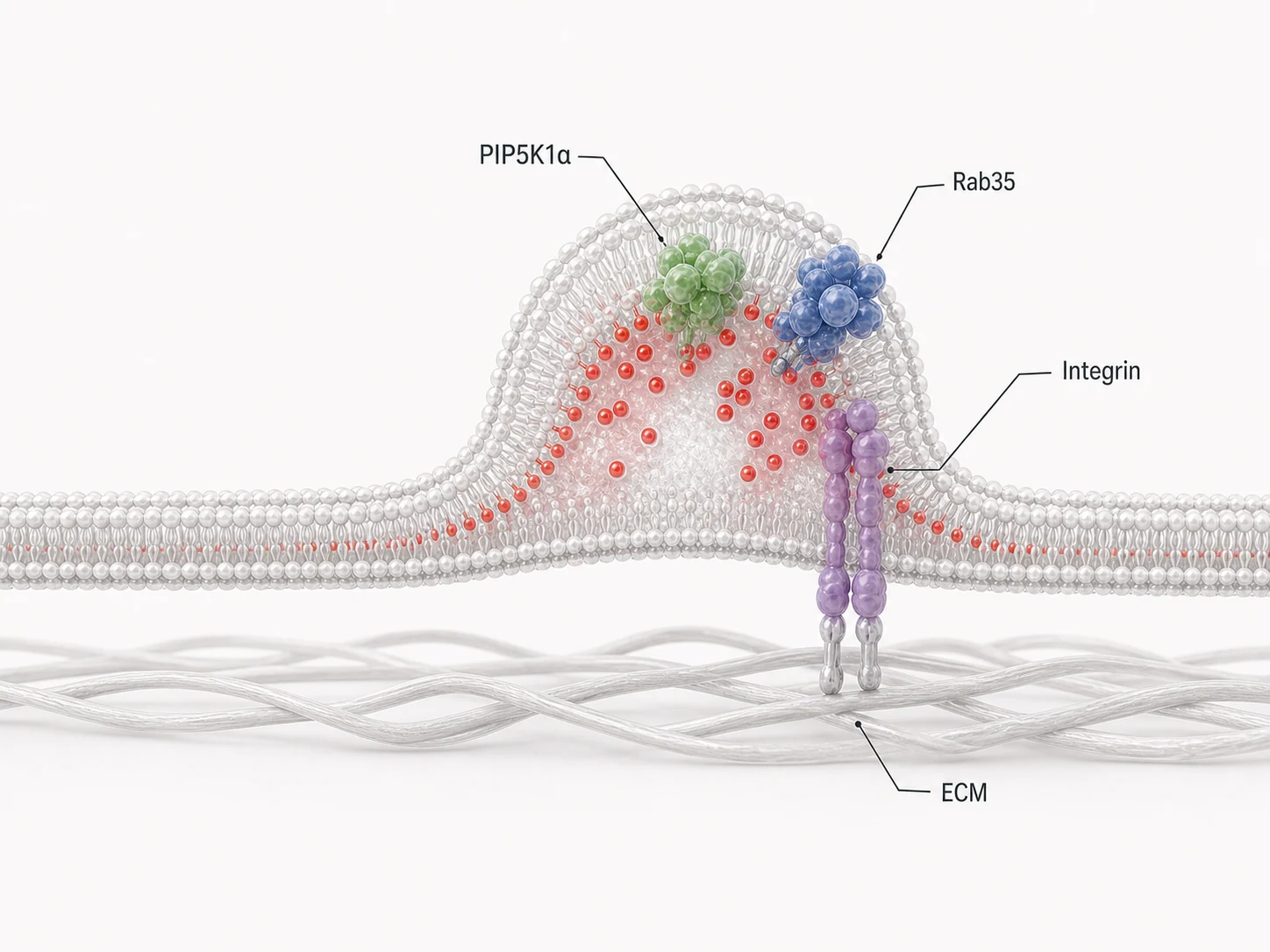
The PIP5K1A-PI(4,5)P2-Rab35-integrin signaling axis establishes mechanically stable formation sites.
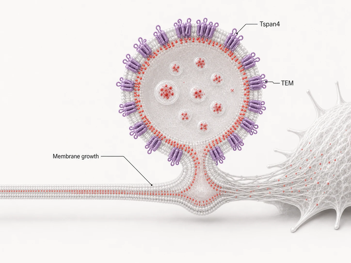
Tetraspanin-enriched membrane domains assemble into larger structures that increase membrane rigidity and stabilize migrasome growth.
02 / WHY
Migrasomes contribute to spatial signaling, cargo transfer and organelle quality control, supporting essential functions across biological systems.
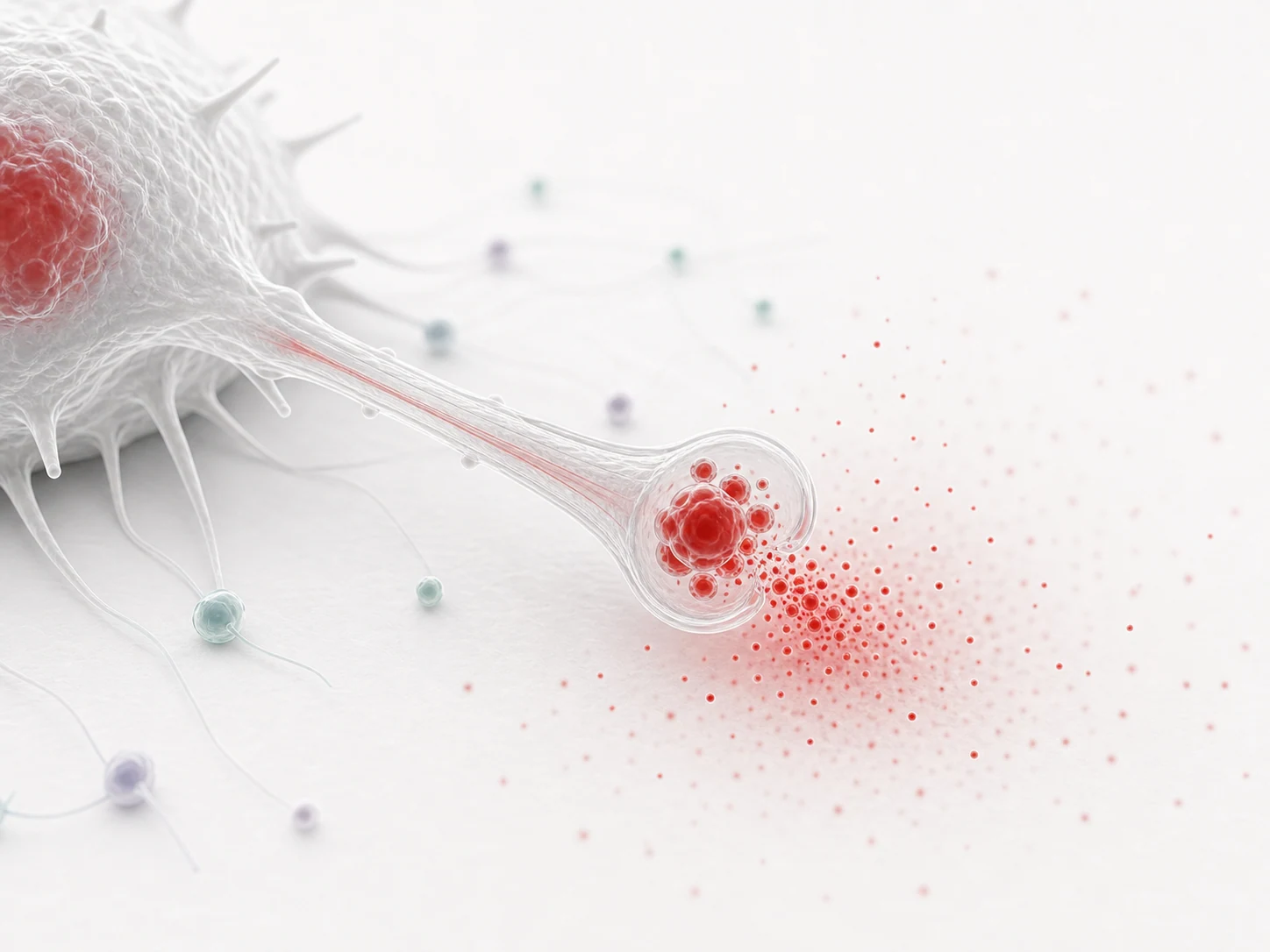
Migrasomes establish localized signaling environments along migration paths, influencing nearby cells.
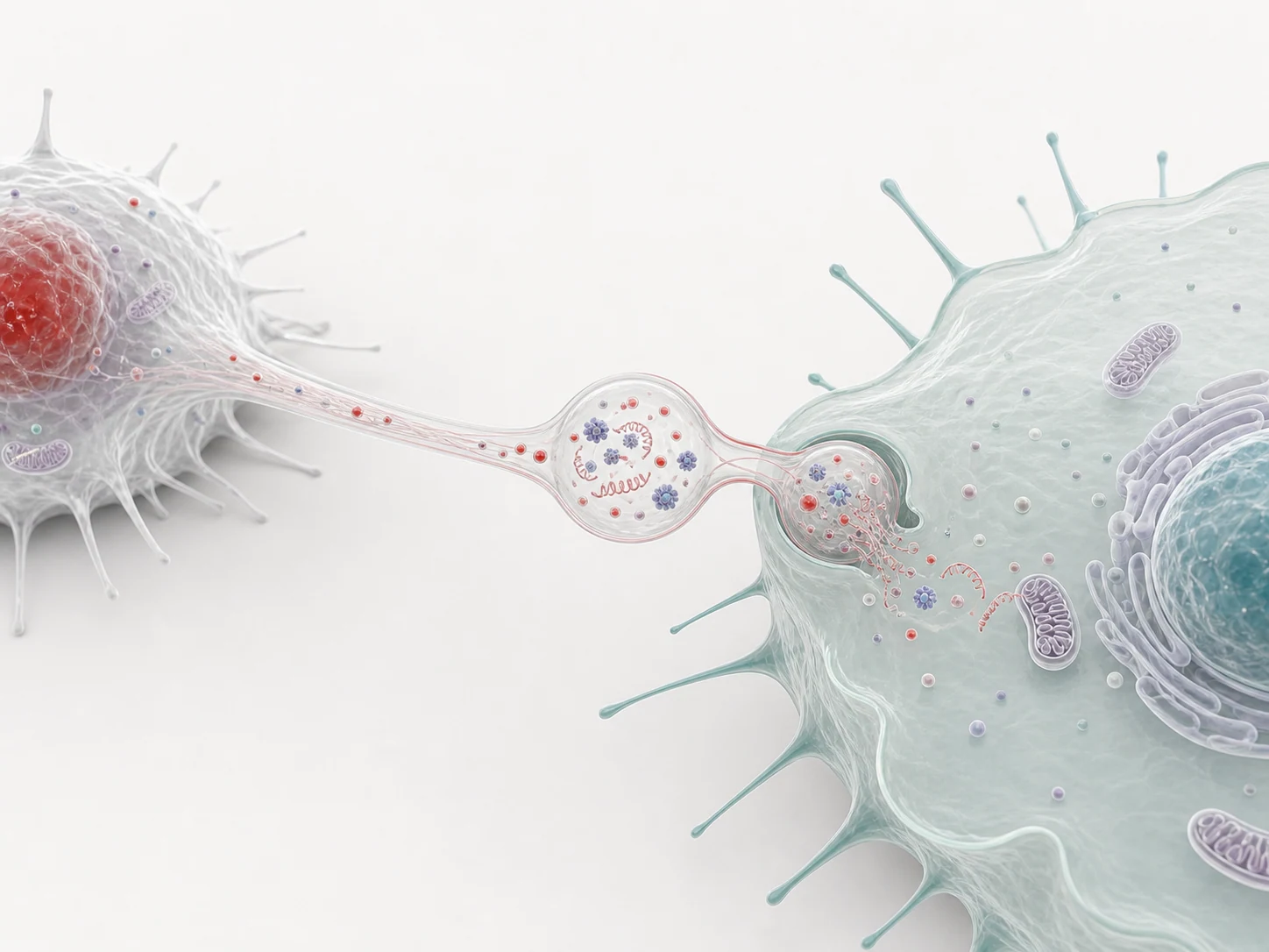
Migrasomes can carry functional cargo, providing a model for material transfer between cells.
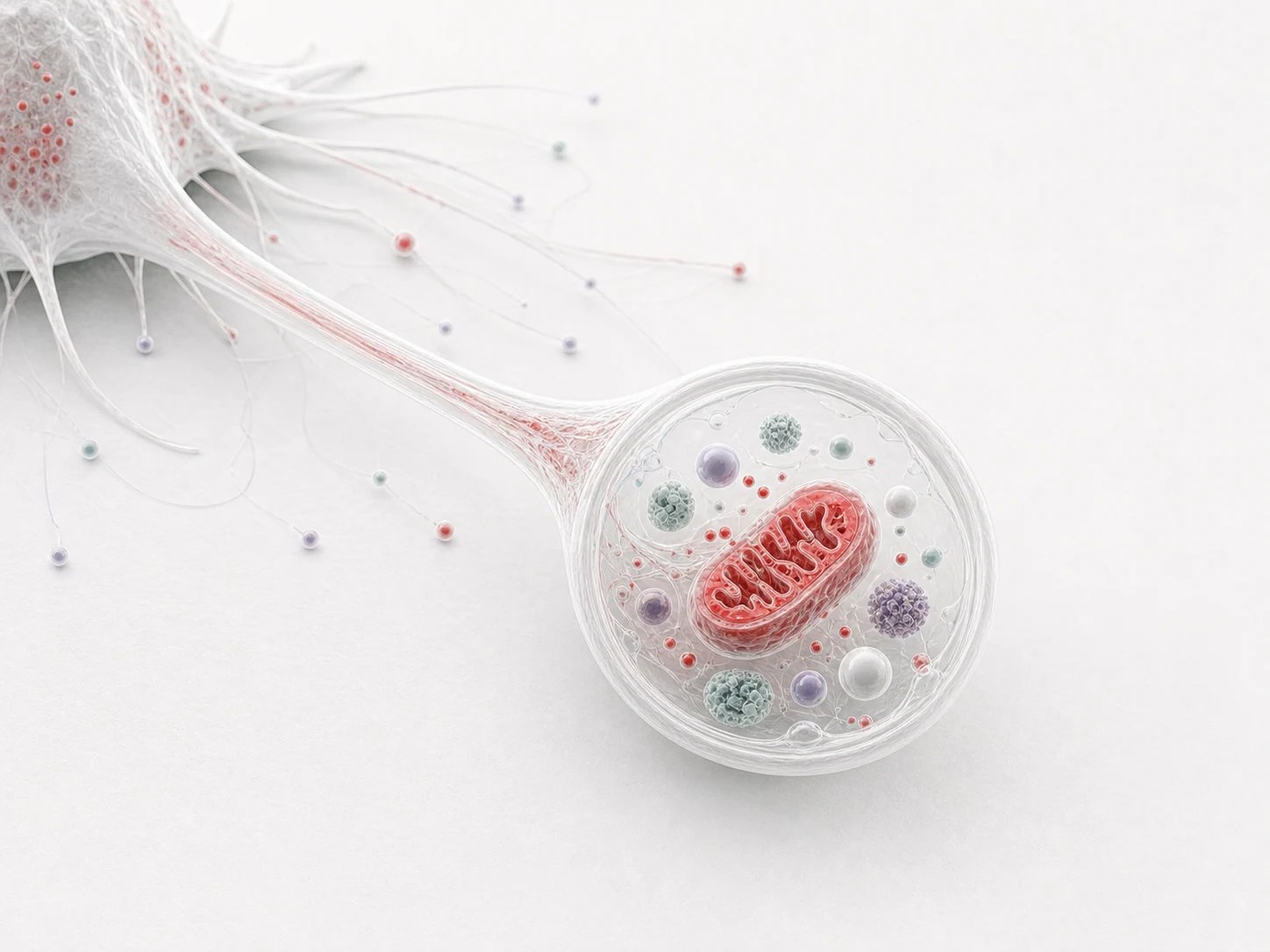
Damaged mitochondrial material can be routed to migrasomes, linking cell movement with cellular homeostasis.
03 / VALUE
Migrasome research is expanding into multiple disease-related fields, creating new directions for mechanism studies, biomarkers and engineered platforms.
Established the concept of migrasomes linked to cell-migration trails.
Showed how extracellular-matrix pairing influences migrasome formation.
Described macro-scale assembly of tetraspanin-enriched domains.
Connected migrasomes with mitochondrial quality control.
Clarified how lipid foci initiate formation sites.

Neutrophil-derived migrasomes were identified as an important component of the coagulation system.
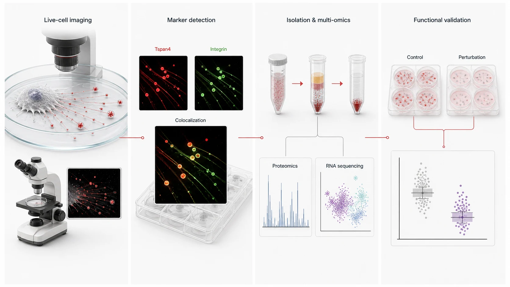
Explore methods, tools, mechanisms, and functions in migrasome research.
04 / HOW
OBSERVE IN SITU
Migrasome formation is closely linked to cell-migration trajectories. High-resolution live-cell imaging can resolve the spatial relationships among migrasomes, retraction fibers and migration paths while recording where and when they form, providing spatial and temporal references for downstream isolation, purification and functional studies.
ISOLATE & PURIFY
Select an appropriate strategy for each sample type. Migrasome Therapeutics provides complete solutions for cultured cells, whole blood/plasma and tissue samples, helping researchers establish reproducible preparation workflows, effectively remove matrix and vesicular impurities, and obtain migrasome samples suitable for downstream analysis.
CHARACTERIZE
Combine morphology, membrane features, markers and cargo composition to connect visible structure with identity and properties.
FUNCTIONAL VALIDATION
Use recipient-cell functional assays, migrasome cargo functional assessment, and genetic or pharmacological perturbations to test causal associations between migrasomes and specific cellular phenotypes.
Functional validation experiments vary by research objective and are typically designed by investigators for their specific studies. The Migrasome technical team can support the full workflow, from experimental design to data interpretation, with practical experience and technical guidance.
Consult the technical team